Dive into the stories of how our solurions have elevated the work of researchers
University of Leeds
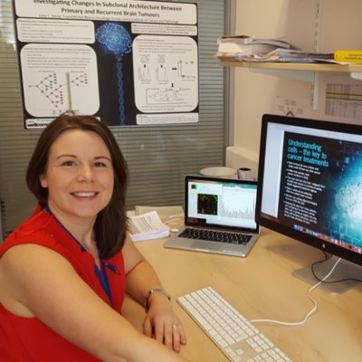
Monitoring ‘Mini Tumours’ growth massed on culture condition and improving knowledge on Gliomodels
We’ve previously used spheroid size as a measure of cell growth under different conditions, but this doesn’t account for cell density so simply isn’t as accurate. The W8 is the first machine that allows us to properly assess how our culture conditions, drugs or different cell lines really impact on the growth of our ‘mini tumours’. The W8 is so much quicker and easier to use than our previous approach of manually sizing our spheroids, and we can even sort them for downstream analysis. We think the W8 is going to be a game-changer for our brain cancer research projects
Dr. Lucy Stead
Associate professor at the University of Leeds and Head of the Gliomodel initiative
Determining Organoid Physical parameters as correlation to cancer aggressiveness in animal models
We have generated organoids from different GEMMs of prostate cancer and observed a plethora of different structures in the cultures system. The W8 has allowed us to associated their density and weights. We have then correlated these physical parameters with different status of aggressiveness of the caner. The W8 is precise instrumentation that will certainly change how we look into new parameters that will allow us to better define the cancer.
IRCCS Istituto Romagnolo per lo Studio dei Tumori
"Dino Amadori" - IRST
Using multidisciplinary approaches to define novel anti-tumor treatments in 3D cell cultures.
Being focused on innovative anti-tumor treatments, with a specific interest in 3D cellular models, some of my goals are related to sample standardization and to the culture of homogenous samples, which are needed to obtain the maximum reliability of the applied treatments. For this reason, I adopt multidisciplinary approaches to grow and characterize 3D spheroids.
In this context, I believe the W8 perfectly fits my research purposes, as it is the only instrument capable to perform the Biophysical analysis, which appeared to be a crucial characterization during the 3D cell models’ standardization. We noted how such biophysical results are giving a novel point to view, not only to highlight sample uniformity, but also to be correlated with the obtained biological results
Digital Microscopy Center - Istituto Ortopedico Rizzoli
Exploring correlations between biophysics and deep imaging in 3D cellular models.
I have been collaborating with CellDynamics since 2017 and have seen the technological level of the group grow exponentially. I believe CellDynamics to be one the most brilliant StartUp and an excellent scientific partner for the development of new investigation techniques on organoids and spheroids.
In recent years we have optimized a complete biophysical analysis of the spheroids that starts from the mass density data obtained with the W8, followed by the sorting of the subpopulations, and then to the clarification of the samples and finally to the microscopic analysis in deep imaging. This workflow allows a precise measurement of the spheroid characteristics and represent a key step forward for the accurate testing of treatment’s potential in 3D in vitro models
We use cookies on our website to give you the most relevant experience by remembering your preferences and repeat visits. By clicking “Accept All”, you consent to the use of ALL the cookies. However, you may visit "Cookie Settings" to provide a controlled consent.
This website uses cookies to improve your experience while you navigate through the website. Out of these, the cookies that are categorized as necessary are stored on your browser as they are essential for the working of basic functionalities of the website. We also use third-party cookies that help us analyze and understand how you use this website. These cookies will be stored in your browser only with your consent. You also have the option to opt-out of these cookies. But opting out of some of these cookies may affect your browsing experience.
Necessary cookies are absolutely essential for the website to function properly. These cookies ensure basic functionalities and security features of the website, anonymously.
Cookie
Duration
Description
cookielawinfo-checkbox-analytics
11 months
This cookie is set by GDPR Cookie Consent plugin. The cookie is used to store the user consent for the cookies in the category "Analytics".
cookielawinfo-checkbox-functional
11 months
The cookie is set by GDPR cookie consent to record the user consent for the cookies in the category "Functional".
cookielawinfo-checkbox-necessary
11 months
This cookie is set by GDPR Cookie Consent plugin. The cookies is used to store the user consent for the cookies in the category "Necessary".
cookielawinfo-checkbox-others
11 months
This cookie is set by GDPR Cookie Consent plugin. The cookie is used to store the user consent for the cookies in the category "Other.
cookielawinfo-checkbox-performance
11 months
This cookie is set by GDPR Cookie Consent plugin. The cookie is used to store the user consent for the cookies in the category "Performance".
viewed_cookie_policy
11 months
The cookie is set by the GDPR Cookie Consent plugin and is used to store whether or not user has consented to the use of cookies. It does not store any personal data.
Functional cookies help to perform certain functionalities like sharing the content of the website on social media platforms, collect feedbacks, and other third-party features.
Performance cookies are used to understand and analyze the key performance indexes of the website which helps in delivering a better user experience for the visitors.
Analytical cookies are used to understand how visitors interact with the website. These cookies help provide information on metrics the number of visitors, bounce rate, traffic source, etc.
Advertisement cookies are used to provide visitors with relevant ads and marketing campaigns. These cookies track visitors across websites and collect information to provide customized ads.